PCR Plate Design and Its Impact on Assay Performance: A Technical Primer for Molecular Biology Laboratories
PCR Plate Design and Its Impact on Assay Performance: A Technical Primer for Molecular Biology Laboratories
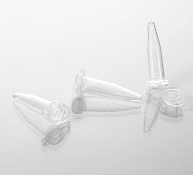
Polymerase chain reaction (PCR) assay development typically focuses on primer design, polymerase selection, and thermal cycling parameters — while the plastic consumable containing the reaction is treated as an inert, interchangeable commodity. Experimental evidence consistently demonstrates otherwise. The geometry, optical properties, and material composition of a PCR plate directly modulate thermal transfer efficiency, fluorescence signal collection, and reaction inhibition — introducing systematic errors that no amount of primer redesign or reference gene normalization can correct.
This article examines four plate design parameters through the lens of their underlying physical mechanisms and their quantitative impact on PCR data quality. The discussion is intended for principal investigators training new laboratory members, graduate students encountering well-to-well variability for the first time, and core facility managers establishing standardized consumables protocols.
1. Well Density and Reaction Volume: Thermal Mass Considerations
The transition from 96-well to 384-well format represents more than a fourfold increase in sample throughput. It fundamentally alters the thermal physics of the reaction environment.
In a 96-well plate with a typical 25 µL reaction volume, the aqueous reaction mixture has a thermal mass that dominates the thermal behavior of the system. The polypropylene well wall (~0.25 mm thickness) acts as a thermal conductor between the aluminum block and the liquid, and the entire liquid volume approaches thermal equilibrium within approximately 3–5 seconds of a block temperature change. This response time is sufficient for standard three-step PCR protocols with 30–60 second step durations.
When reaction volume is reduced to 5 µL for 384-well compatibility, the thermal mass decreases proportionally — but so does the tolerance for pipetting error. At 5 µL total volume, a ±0.2 µL pipetting deviation (well within the specification of most calibrated air-displacement pipettes) represents a ±4% volume error. The same absolute error at 25 µL is ±0.8%. This error propagates non-linearly into Ct values: for a target present at 1,000 copies per reaction, a 4% volume reduction concentrates the template, producing a Ct shift of approximately 0.06 cycles — negligible in isolation but cumulatively significant when combined with other sources of technical variation across a 384-well plate.
Evaporation kinetics introduce a second volume-dependent artifact. Water vapor loss from a well during thermal cycling proceeds at a rate proportional to the liquid surface area and the vapor pressure gradient between the liquid and the headspace. A 5 µL reaction in a 384-well plate has a surface-area-to-volume ratio approximately 1.6× higher than a 25 µL reaction in a 96-well plate. Under identical sealing conditions, the 384-well reaction loses a larger fraction of its total volume to evaporation over 40 cycles. The resulting solute concentration increases fluorophore signal intensity, producing artificially lower Ct values in affected wells — an effect that is indistinguishable from genuine template abundance differences without running a standard curve on every plate.
Practical guidance: For laboratories processing fewer than 200 samples per day, 96-well plates with 20–25 µL reaction volumes provide the optimal balance of pipetting precision and reagent economy. The transition to 384-well format should coincide with the introduction of automated liquid handling; manual 384-well pipetting introduces well-documented cross-contamination risks from the 4.5 mm inter-well spacing that typically exceed any reagent cost savings.
Individual PCR Tube Formats: 0.1, 0.2, and 0.5 mL
While plate-based formats dominate high-throughput workflows, individual PCR tubes remain essential for low- to medium-throughput applications, protocol development, and laboratories that process variable sample numbers. The three standard tube volumes — 0.1 mL, 0.2 mL, and 0.5 mL — are not interchangeable; each is optimized for specific thermal cycler block geometries and reaction volume ranges.
| Parameter | 0.1 mL Tube | 0.2 mL Tube | 0.5 mL Tube |
|---|---|---|---|
| Typical reaction volume | 5–30 µL | 10–100 µL | 25–200 µL |
| Maximum volume | ~50 µL | ~150 µL | ~300 µL |
| Wall thickness | Ultra-thin (~0.15–0.20 mm) | Thin (~0.20–0.25 mm) | Thin to standard (~0.25–0.30 mm) |
| Thermal cycler compatibility | Fast/ultra-fast cyclers; 96-well low-profile blocks; some qPCR instruments with 0.1 mL-specific blocks | Standard 96-well blocks — the most universal format; compatible with virtually all modern thermal cyclers | Older cycler models with 0.5 mL blocks; some specialized clinical protocols; preparative PCR |
| Thermal equilibration | Fastest — minimal thermal lag; ideal for fast PCR (<10 s/step) | Fast — good balance of speed and reaction volume flexibility | Moderate — larger thermal mass; longer equilibration time |
| Evaporation risk | Higher — small absolute volume loss is a large fractional loss; good sealing essential | Moderate — typical for most protocols | Lower — larger volume buffer against evaporative loss |
| Best application | Fast qPCR, low-volume reactions, precious templates, ultra-fast cycling protocols | Standard qPCR, conventional PCR, genotyping — the default choice for most research laboratories | Preparative PCR, large-volume reactions, clinical protocols on older instrumentation, sample archiving |
| Cap style | Flat or domed; flat caps preferred for optical qPCR (minimal light scattering) | Flat or domed; flat caps enable labeling; domed caps provide marginally better seal | Flat cap standard; secure seal for long-term storage of amplified product |
The 0.2 mL tube is the de facto standard for most academic and clinical research laboratories. It is the format supported by the broadest range of thermal cycler models and strikes the optimal balance between reaction volume flexibility, thermal performance, and reagent economy for typical 20–50 µL reactions. The 0.1 mL format is gaining adoption in qPCR-centric laboratories where fast cycling and minimal reagent consumption are priorities; however, careful attention to sealing is required due to the high surface-area-to-volume ratio and consequent evaporation sensitivity. MUHWA supplies certified PCR tubes across all three volume formats: 0.1 mL and 0.2 mL in a combined pack (0.1/0.2mL PCR Tubes with Flat Cap, 1000/pack) for standard qPCR and conventional PCR workflows, and 0.5 mL (0.5mL Thin-Wall PCR Tubes, 1000/pack) for preparative PCR and larger-volume clinical protocols. All tubes are manufactured from virgin polypropylene with batch-level quality control.
When transitioning from individual tubes to 96-well plates, note that plate wells are functionally equivalent to 0.2 mL tubes in thermal mass and volume capacity. Low-profile plate wells approximate 0.1 mL tube performance characteristics. The same principles of tube material quality — virgin polypropylene, certified nuclease-free — apply equally to individual tubes and multi-well plates.
Tube Strips: 8-Tube and 12-Tube Formats
For laboratories that process samples in multiples of 8 or 12 and wish to avoid the per-tube handling overhead of individual tubes without committing to a full 96-well plate, PCR tube strips provide an efficient intermediate format. Strips are manufactured in two standard configurations: 8-tube strips (compatible with 8-channel multichannel pipettes, matching one column of a 96-well plate) and 12-tube strips (compatible with 12-channel pipettes, matching one row).
| Property | Individual Tubes | 8-Tube Strips | 12-Tube Strips | 96-Well Plate |
|---|---|---|---|---|
| Sample flexibility | Maximum — run any number (1–96) | Moderate — must run in multiples of 8 | Moderate — must run in multiples of 12 | Minimum — entire plate consumed; wasted wells for small batches |
| Setup speed | Slow — cap each tube individually | Fast — 8-cap strip seals all tubes at once | Fast — 12-cap strip seals all tubes at once | Fastest — adhesive film or heat seal covers 96 wells simultaneously |
| Sample tracking | Risk of mislabeling or tube mix-up | Fixed order within strip reduces mix-up; strip can be labeled as a unit | Same as 8-tube strip | Well position is fixed; plate barcode enables automated tracking |
| Multichannel pipette | Compatible but alignment is manual | Designed for 8-channel pipettes — pitch matches perfectly | Designed for 12-channel pipettes | Compatible with both 8- and 12-channel |
| Well recovery | Individual tubes can be removed and processed separately | Individual tubes can be cut from strip with scissors if needed | Same — tubes can be separated | Individual wells cannot be physically separated |
| Cost per sample | Moderate | Slightly lower than individual tubes | Slightly lower than individual tubes | Lowest per well at high throughput |
| Best application | Variable sample numbers, protocol development, low throughput | Medium-throughput qPCR; genotyping panels; reactions aligned with 8-channel pipetting workflow | Medium-throughput protocols using 12-channel pipettes; row-based sample organization | High-throughput screening; clinical diagnostic batches; automated liquid handling |
The 8-tube strip is the most commonly used strip format because 8-channel multichannel pipettes are standard equipment in most molecular biology laboratories. The 8-well format maps directly to one column of a 96-well plate, making strips the natural choice when a full plate is unnecessary but the multichannel pipetting workflow is desired. For laboratories running 20–40 samples per batch — too many to cap individually, too few to justify a full plate — 8-tube strips in a 96-well rack deliver the speed of plate-based pipetting with the flexibility of individual tubes. MUHWA offers 0.1 mL 8-strip PCR tubes with flat caps (125 strips/pack), manufactured from clear virgin polypropylene with thin-wall construction for rapid thermal transfer in qPCR and fast PCR protocols.
2. Skirt Geometry: Thermal Expansion, Mechanical Constraint, and Block Contact Uniformity
The skirt — a polypropylene frame extending below the plane of the well bottoms — is often regarded as a mechanical convenience for plate handling. Its primary function is structural: resisting thermal deformation during repeated temperature cycling.
The mechanism is straightforward but underappreciated. Polypropylene and aluminum have markedly different coefficients of thermal expansion (CTE): approximately 100–150 × 10⁻⁶ /K for polypropylene versus 23 × 10⁻⁶ /K for the aluminum alloy typically used in cycler blocks. During a temperature ramp from 60°C (annealing/extension) to 95°C (denaturation), a 127 mm polypropylene plate expands by approximately 0.4–0.6 mm relative to the aluminum block. This differential expansion generates internal mechanical stress within the plate, concentrated at the points of constraint — the wells seated in the block.
In an unskirted plate, the entire structure deforms to relieve this stress, producing a concave bow that develops progressively over 30–40 thermal cycles. The resulting air gap between affected wells and the block surface introduces thermal contact resistance. Since air has a thermal conductivity of approximately 0.026 W/m·K — roughly four orders of magnitude lower than aluminum (205 W/m·K) — even a 0.1 mm air gap creates a significant thermal barrier. Wells in the center of the plate, where bowing is maximal, may operate 1–3°C below the block setpoint during the denaturation step, compromising strand separation efficiency for GC-rich templates.
Semi-skirted plates constrain the plate along two edges, reducing but not eliminating warping. The residual deformation is typically asymmetric, producing a thermal gradient across the plate that correlates with well position — a systematic error that reference gene normalization cannot correct, because it affects all targets in affected wells equally.
Full-skirted plates constrain all four edges, providing the mechanical rigidity to maintain planarity through repeated cycling. The continuous sealing surface around the entire perimeter also ensures uniform adhesive film contact, directly reducing the edge-well evaporation that produces the well-known "edge effect" — systematically elevated Ct values in columns 1 and 12, rows A and H.
Quantitative expectation: Well-position-dependent Ct variation in unskirted plates typically ranges from 0.5–1.5 cycles across a 96-well plate after 40 cycles. Full-skirted plates reduce this to 0.1–0.3 cycles — within the range of stochastic variation for most assays. For quantitative PCR where ±0.5 Ct represents the threshold for biological significance, this difference is dispositive.
3. Plate Color and Fluorescence Detection Physics
The optical specification of a PCR plate directly governs signal collection efficiency in real-time fluorescence detection systems. This is not a question of plate aesthetics — it is governed by the geometry of fluorescence emission and the optical path of the detection system.
Fluorescence emission from an excited fluorophore (SYBR Green I, TaqMan probe, molecular beacon) is isotropic — photons are emitted in all directions with equal probability. In a transparent (clear) plate, photons emitted laterally pass through the well wall into the adjacent well, where they are detected as spurious signal. This optical crosstalk elevates the apparent baseline fluorescence in neighboring wells, compressing the dynamic range at low template concentrations and generating false-positive signals in no-template control (NTC) wells adjacent to high-template samples.
The magnitude of this effect depends on the specific detection geometry. Instruments employing top-reading optics (Roche LightCycler 480, Bio-Rad CFX series) are more susceptible to inter-well crosstalk from clear plates because the detector collects photons from above the plate, where scattered and reflected light from adjacent wells contributes to the signal. Bottom-reading instruments (older ABI 7900HT configurations) show reduced but not eliminated crosstalk, as the excitation beam necessarily passes through the plate material.
White plates address both mechanisms simultaneously. Titanium dioxide (TiO₂) pigment dispersed in the polypropylene matrix creates optically reflective well walls. Fluorescence emission that would otherwise escape laterally is reflected internally toward the detector — increasing photon collection efficiency by approximately 2–3× relative to clear plates, depending on detector geometry. The opaque walls physically block inter-well photon transmission, effectively eliminating optical crosstalk. The combined effect is a measurable improvement in signal-to-background ratio, with typical Ct improvements of 0.5–1.0 cycles for low-copy-number targets and tighter technical replicate CVs.
The practical significance is most apparent at the extremes of the dynamic range. For a target present at 10–100 copies per reaction — where stochastic variation already dominates — the signal improvement from white plates can mean the difference between detection and non-detection. For high-template samples, the crosstalk reduction eliminates the characteristic "ramping" of NTC wells in columns adjacent to strong positive samples — a troubleshooting finding that otherwise suggests contamination when none exists.
4. Well Profile: Heat Transfer Kinetics
The depth of a PCR plate well determines the height of the liquid column and, consequently, the time required for the entire reaction volume to reach thermal equilibrium following a block temperature change.
Heat transfer from the cycler block into the reaction volume proceeds primarily by conduction through the polypropylene well bottom, followed by convective mixing within the liquid. The rate-limiting step is conduction through the liquid itself: water has a thermal diffusivity of approximately 1.4 × 10⁻⁷ m²/s, meaning that a 15 mm liquid column (standard-profile well) requires approximately 3–5 seconds to equilibrate after a rapid block temperature change, while a 10 mm column (low-profile well) equilibrates in 1–2 seconds.
During fast PCR protocols — increasingly common as instrument ramp rates exceed 4°C/second — this equilibration lag produces a transient vertical temperature gradient within the well. The liquid at the bottom of the well, in direct contact with the heated polypropylene surface, may be 2–3°C warmer than the liquid at the top during the initial seconds of a denaturation step. If fluorescence acquisition occurs during this transient period (as it does in many fast qPCR protocols), the measured signal reflects conditions in only a fraction of the reaction volume.
Low-profile plates reduce this gradient by shortening the thermal path. For qPCR applications with rapid cycling, the improvement in temperature uniformity at the acquisition timepoint translates to tighter technical replicates and improved assay reproducibility. A recent multi-laboratory study (Vandesompele et al., 2021; Clinical Chemistry) demonstrated that low-profile plates reduced inter-well Ct standard deviation by approximately 15–20% compared to standard-profile plates under fast cycling conditions (>4°C/s ramp rate, <10 s step duration).
The trade-off is well capacity: low-profile plates typically accommodate 5–50 µL, while standard-profile plates accept 15–200 µL. For preparative PCR applications requiring large product volumes, standard-profile plates remain appropriate.
A critical note on compatibility: Using a low-profile plate in a cycler with a standard-profile block creates a 3–5 mm air gap below the well bottoms. The resulting thermal barrier produces uniform but systematic Ct delays of 1–3 cycles — easily mistaken for reduced template abundance. Conversely, a standard-profile plate in a low-profile block may not seat fully, risking mechanical damage to both plate and instrument. The well profile must match the instrument specification.
5. Polymer Purity: The Underappreciated Variable in PCR Inhibition
Polypropylene is the universal material for PCR consumables because it satisfies three essential requirements: thermal stability to >120°C, chemical inertness toward common PCR buffer components, and low non-specific binding of nucleic acids and proteins. These properties, however, apply to pure polypropylene — and the distinction between virgin resin and recycled or blended feedstock is functionally significant.
Recycled polypropylene originates from post-industrial or post-consumer waste streams and necessarily contains residues from its previous manufacturing and use history. Common contaminants include phthalate and adipate plasticizers (added to improve flexibility in consumer products), fatty acid ester mold-release agents (used in injection molding), and thermal-oxidative degradation products from repeated melt-processing (carbonyl compounds, hydroperoxides). These compounds are lipophilic, thermally stable, and not removed by standard regrind-and-repelletize recycling processes.
When introduced into a PCR reaction and subjected to repeated heating to 95°C, these leachates inhibit Taq DNA polymerase — the mechanistic basis appears to involve competitive binding at the enzyme's hydrophobic active-site cleft or non-specific interaction with the template DNA. The inhibition is partial: reactions still amplify, but with reduced efficiency. A 2018 comparative study (Murray et al., BioTechniques) documented amplification efficiency reductions of 25–40% in reactions performed in recycled-resin tubes versus virgin polypropylene controls, as measured by standard curve slope deviation.
Critically, this inhibition is not uniform across all wells or all plates — it varies with the spatial distribution of contaminants within the injection-molded part. The resulting well-to-well variation in amplification efficiency is indistinguishable from genuine biological or stochastic variation, and no amount of reference gene normalization can correct for differential inhibition across wells.
Nuclease contamination introduces a separate but equally consequential failure mode. RNase A, the predominant environmental ribonuclease, is a remarkably stable enzyme that survives autoclaving at 121°C and renatures upon cooling. A single fingerprint transfers sufficient RNase to degrade microgram quantities of RNA. For RT-qPCR workflows, RNase contamination in the plate well — introduced during manufacturing, packaging, or handling — degrades the RNA template before reverse transcription initiates, producing truncated cDNA and inconsistent amplification that may be misattributed to poor RNA quality or suboptimal reverse transcription conditions.
The certification standard matters. Batch-level RNase/DNase testing using validated fluorometric nuclease assays (detection limit ~10⁻⁶ Kunitz units) with lot-specific certificates of analysis represents current best practice. Vague manufacturer claims of "clean manufacturing" without quantitative testing data should be treated as functionally equivalent to uncertified product.
6. Sealing Methods: Evaporation, Condensation, and the Edge-Well Problem
Well sealing is the most operator-dependent variable in PCR plate preparation — and the largest preventable source of well-to-well Ct variation. The underlying physical process is straightforward: water evaporates from the heated reaction volume, condenses on the cooler underside of the seal or lid, and either remains as droplets (reducing headspace humidity and accelerating further evaporation) or returns to the reaction volume (introducing thermal non-uniformity).
Edge wells are disproportionately affected because their seals experience asymmetric mechanical constraint: the adhesive film at the plate perimeter has only one neighboring well on one side, reducing the effective bonding area and increasing the probability of micro-leaks. This is why columns 1 and 12, rows A and H consistently show higher Ct values in qPCR plates — not because those wells contain less template, but because they contain less water by the end of the run.
Adhesive films (pressure-sensitive acrylic or silicone adhesives on transparent polyester or polypropylene backing) are the most widely used sealing method. When applied with a hand roller, seal quality depends on operator technique: insufficient pressure, especially at the plate perimeter, leaves micro-channels that permit evaporation. A pressure applicator ($500–1,500) standardizes the sealing force and improves edge-well consistency, though the capital cost is only justified in high-throughput settings.
Heat sealing eliminates the adhesive variable entirely by melting a polymer film directly into the plate surface, forming a fusion bond. The resulting seal is essentially evaporation-proof — volume loss <0.5% over 40 cycles — and remains intact during frozen storage, making heat-sealed plates the only format validated for long-term archiving of amplified product. The capital equipment cost ($2,000–4,000) is the primary barrier to adoption in academic laboratories; core facilities that serve multiple research groups frequently provide heat sealing as a shared service.
Cap strips (8-cap or 12-cap strip formats) offer the advantage of individual well access post-amplification — useful when PCR product will be recovered for downstream cloning or sequencing. However, domed caps can mechanically interfere with heated lids in some cycler designs, and the per-well cost is higher than film-based sealing.
7. Summary of Recommendations by Application
| Application | Format | Skirt | Color | Profile | Seal |
|---|---|---|---|---|---|
| Standard PCR → gel electrophoresis | 96-well | Semi-skirt | Clear | Standard | Adhesive film |
| qPCR — gene expression (SYBR Green) | 96-well | Full skirt | White | Low-profile | Adhesive film (roller) or heat seal |
| qPCR — clinical assay (TaqMan probe) | 96-well | Full skirt | White | Low-profile | Heat seal (validated) |
| RT-qPCR — RNA template | 96-well | Full skirt | White | Low-profile | Adhesive film or heat seal |
| High-throughput screening | 384-well | Full skirt | White | Low-profile | Heat seal |
| SNP genotyping (endpoint) | 96 or 384 | Full skirt | White | Low-profile | Adhesive film |
| Preparative PCR (product recovery) | 96-well | Semi-skirt | Clear | Standard | Cap strips |
This technical reference is provided for educational purposes. The quantitative performance characteristics discussed are derived from published peer-reviewed literature and manufacturer validation data; individual results may vary with specific instrument models, protocol parameters, and environmental conditions. Clinical laboratories should validate all consumables per their quality management system prior to diagnostic use.
#PCR plate #qPCR optimization #PCR consumables #molecular biology methods #laboratory techniques #thermal cycling #fluorescence detection #PCR troubleshooting #experimental design #graduate education
Comments
Post a Comment