PCR Contamination Control: Sources, Mechanisms, and Laboratory Protocols for Preventing False-Positive Amplification
PCR Contamination Control: Sources, Mechanisms, and Laboratory Protocols for Preventing False-Positive Amplification
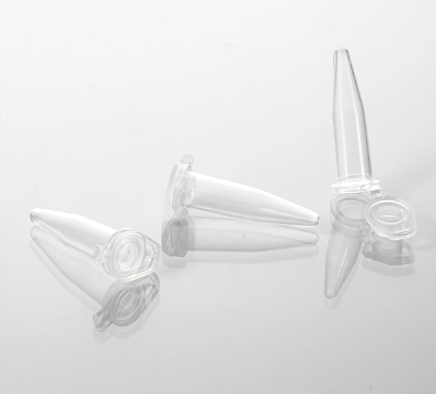
PCR contamination is a laboratory problem with a distinctive and frustrating characteristic: it is usually recognized only after it has already compromised experimental data. A no-template control (NTC) that produces a positive signal may represent a single contaminated reagent aliquot — or it may indicate that aerosolized amplicons from previous reactions have distributed throughout the laboratory environment, contaminating surfaces, equipment, and reagent stocks in a pattern that requires systematic decontamination rather than simple reagent replacement. Distinguishing between these scenarios — and implementing preventive measures that make the distinction unnecessary — is a core competency for laboratories performing amplification-based assays.
This article examines the physical and biochemical mechanisms of PCR contamination, the rationale for spatially segregated workflow design, and the control reactions that provide early detection of contamination events before they propagate through multiple experiments. The protocols described reflect current best practice in clinical molecular diagnostics and have been adapted for academic research laboratory settings where dedicated physically separated rooms may not be available.
1. Contamination Sources: Aerosols, Surfaces, and Reagent Pathways
PCR contamination originates from three mechanistically distinct sources, each requiring different preventive strategies.
1.1 Aerosolized Amplicons
After 30–40 cycles of amplification, a single PCR tube contains approximately 10¹²–10¹³ copies of the target amplicon. Opening this tube — for gel loading, sequencing, or product purification — releases an aerosol of microscopic droplets, each potentially containing thousands of amplifiable DNA molecules. These droplets, typically 0.5–10 µm in diameter, remain suspended in air for minutes to hours and settle onto all exposed surfaces within the workspace.
The contamination potential is extraordinary: a single 1 µL droplet from a completed PCR reaction contains approximately 10⁹–10¹⁰ amplicon copies. The typical detection limit of a well-optimized qPCR assay is 1–10 copies per reaction. This represents a contamination-to-detection ratio of approximately 10⁹:1 — meaning that even a droplet volume of 10⁻⁹ µL (one femtoliter, far below visual detection) contains sufficient template to produce a strong positive signal. This is why aerosol contamination, once established, can persist for weeks despite routine cleaning: the contaminating template is present at concentrations that are functionally undetectable by any method other than PCR itself.
The physical distribution of aerosol contamination follows airflow patterns within the laboratory. Laminar flow hoods and biosafety cabinets direct aerosols away from the operator and sample, but standard laboratory benches without directional airflow allow aerosols to accumulate on all nearby surfaces — pipettes, tube racks, reagent bottles, notebooks, and personal items including phones and pens carried between laboratory areas.
1.2 Surface-Mediated Cross-Contamination
Direct transfer of template DNA between samples occurs through contact with contaminated surfaces. Pipette tips that touch the rim of a template-containing tube transfer nanogram quantities of DNA to the next tube contacted. Gloves that handle template tubes, then touch reagent bottle caps, transfer template to the reagent stock — from which it is subsequently aliquoted into every reaction prepared from that bottle.
The persistence of dried DNA on laboratory surfaces is considerable. Double-stranded DNA adsorbed onto polystyrene, polypropylene, or stainless steel surfaces remains amplifiable for weeks under standard laboratory conditions (22°C, ambient humidity). Desiccation does not degrade the template; it merely reduces the hydration sphere, which is restored upon contact with aqueous PCR master mix. Autoclaving inactivates biological organisms but does not destroy DNA — the phosphodiester backbone is chemically stable at 121°C, and depurination, while accelerated at elevated temperature, proceeds slowly enough that amplifiable fragments of >100 bp routinely survive standard autoclave cycles.
1.3 Reagent Contamination
Reagent stocks — master mix, primer solutions, PCR-grade water, MgCl₂ — become contaminated when a pipette tip that has contacted template DNA is inserted into the stock bottle. Because these stocks are used to prepare multiple batches of reactions, a single contamination event propagates through all subsequent experiments using that reagent batch. The resulting false-positive signals are consistent across plates and across days — a pattern that distinguishes reagent contamination from sporadic surface or aerosol contamination, which typically produces intermittent positives.
Commercial master mixes are supplied as sterile, DNA-free solutions, but they are not sterile after opening. Each insertion of a pipette tip into the bottle introduces a contamination opportunity. The cumulative probability of contamination increases with the number of accesses, making large shared stock bottles particularly high-risk in multi-user laboratories.
2. Spatial Workflow Design: The Unidirectional Principle
The fundamental principle of PCR contamination control is unidirectional workflow: materials, equipment, and personnel move from clean (pre-amplification) to dirty (post-amplification) areas, and never in the reverse direction. This principle is implemented through physical separation, dedicated equipment, and procedural rules.
2.1 Three-Zone Laboratory Design
Clinical molecular diagnostics laboratories — where false-positive PCR results have direct patient-care consequences — employ physically separated rooms for each workflow stage. Research laboratories with limited space can implement functional equivalents within a single room by designating distinct work areas and maintaining strict unidirectional workflow discipline.
| Zone | Function | Dedicated Equipment | Prohibited |
|---|---|---|---|
| Zone 1: Reagent Preparation | Master mix assembly, reagent aliquoting, preparation of NTC reactions | Pipettes (set A), filter tips, tubes, ice bucket, microcentrifuge, vortex | Template DNA of any kind; amplified PCR product; plasmids; genomic DNA extracts |
| Zone 2: Template Addition | Addition of DNA/cDNA template to prepared master mix; tube sealing | Pipettes (set B), filter tips, tube racks, dedicated glove box if available | Amplified PCR product; movement of materials back to Zone 1 |
| Zone 3: Amplification & Post-PCR Analysis | Thermal cycling, gel electrophoresis, product purification, sequencing | Thermocycler, gel electrophoresis equipment, power supply, imaging system | Entry into Zone 1 or 2 without glove change, lab coat change, and surface decontamination |
The spatial order matters: Zone 1 should be positioned farthest from Zone 3, with Zone 2 in between. In a single-room implementation, this translates to a left-to-right workflow along the bench, with clearly demarcated boundaries between zones. The reverse path — Zone 3 to Zone 1 — should require a deliberate decontamination procedure (glove change, surface wipe, if possible lab coat change) that enforces the unidirectional principle by making violations procedurally inconvenient.
2.2 Dedicated Equipment and Color Coding
Pipettes are the primary vector for cross-zone contamination. Each zone requires its own pipette set, clearly labeled and never transferred between zones. A color-coding system (e.g., green tape = Zone 1 reagent prep, yellow = Zone 2 template addition, red = Zone 3 post-PCR) provides immediate visual confirmation of proper equipment placement. The modest cost of dedicated pipette sets ($200–500 per set for research-grade pipettes) is negligible compared to the cost of repeating experiments compromised by contamination.
Ice buckets, tube racks, microcentrifuges, and vortex mixers should similarly be zone-dedicated. If equipment sharing is unavoidable (e.g., a single microcentrifuge serving multiple zones), strict decontamination — 10% bleach wipe followed by 70% ethanol — must precede each zone transition. In practice, the procedural friction of shared equipment decontamination inevitably leads to compliance failures; dedicated equipment is the more reliable strategy.
3. Reagent Management: Aliquoting and Single-Use Principles
The most effective single intervention for preventing reagent contamination is aliquoting upon receipt: immediately dividing commercial reagent stocks into single-use or limited-use volumes in sterile, DNA-free tubes. This practice isolates contamination events: if one aliquot becomes contaminated, only reactions prepared from that aliquot are affected, and the remaining aliquots in frozen storage remain pristine.
The economic case is straightforward. A 5 mL bottle of 2× qPCR master mix ($200–400) prepares approximately 400 reactions at 25 µL each. A single contamination event — one pipette tip that previously contacted template DNA — renders the entire 5 mL stock unreliable. The cost of 20 sterile microcentrifuge tubes for aliquoting ($5–10) represents approximately 2–5% of the stock value and eliminates the single-point-of-failure risk.
UV decontamination of master mix is employed in some laboratories but requires careful consideration of its limitations. UV-C irradiation (254 nm) induces cyclobutane pyrimidine dimers and 6-4 photoproducts in contaminating DNA, rendering it unamplifiable. However, UV penetrates aqueous solutions poorly — the absorption coefficient of water at 254 nm is approximately 0.01 cm⁻¹, meaning that a 1 cm path length attenuates the incident intensity by only ~2%, but dissolved organics and particulates dramatically increase effective absorbance. In practice, UV treatment is surface-limited: it decontaminates the air-liquid interface of a master mix aliquot but does not reliably penetrate the full volume. It also damages Taq polymerase if the enzyme is present during irradiation — UV must be applied to the master mix before enzyme addition, which adds a procedural step and a contamination opportunity. Aliquot discipline is more reliable and requires no specialized equipment.
4. Control Reactions: Design, Interpretation, and Troubleshooting
Control reactions are the diagnostic system for PCR contamination. Each control type interrogates a specific contamination pathway, and the pattern of control failures identifies the contamination source.
| Control | Composition | Expected Result | Failure Indicates |
|---|---|---|---|
| No-Template Control (NTC) | Master mix + primers + PCR-grade water (no template) | No amplification | Contamination in master mix, water, primers, or plate/tube. The most informative control — run at least 2 NTCs per plate, distributed across plate positions (not adjacent). |
| Positive Control | Master mix + primers + known positive template at defined concentration | Amplification at expected Ct (±1 cycle of historical average) | Reagent failure, thermal cycler malfunction, or pipetting error. A failed positive control invalidates negative results (false negatives). |
| Extraction Negative Control | Blank sample (nuclease-free water or empty tube) carried through entire DNA/RNA extraction procedure, then used as template | No amplification | Contamination introduced during nucleic acid extraction — contaminated extraction reagents, columns, or consumables. This control distinguishes pre-PCR contamination from extraction-phase contamination. |
| No-RT Control (RT-qPCR only) | RNA template subjected to reverse transcription master mix without reverse transcriptase enzyme | No amplification | Genomic DNA contamination in RNA sample. Essential for RT-qPCR; a positive no-RT control indicates that the signal originates from DNA, not cDNA. |
Interpreting NTC failure: A positive NTC demands immediate investigation — do not report sample results from that plate. The diagnostic sequence is: (1) repeat the NTC with fresh aliquots of each individual reagent component to isolate the contaminated stock, (2) if fresh aliquots resolve the NTC, discard the contaminated aliquot and resume work, (3) if fresh aliquots do not resolve the NTC, surface or aerosol contamination is present and full decontamination is required. Laboratories that run NTCs sporadically rather than on every plate lose the temporal resolution needed for this diagnostic sequence.
5. Decontamination Chemistry and Protocol
When contamination is confirmed — persistent NTC positivity after reagent replacement — systematic decontamination is required. The chemistry is straightforward but sequence-dependent.
5.1 Bleach (Sodium Hypochlorite) Oxidation
Sodium hypochlorite (NaOCl, commercial bleach at 5–6% w/v, diluted to 10% v/v working solution ≈ 0.5–0.6% NaOCl) oxidizes the deoxyribose sugar-phosphate backbone of DNA, fragmenting the polymer into short oligomers that cannot serve as PCR templates. The reaction is rapid — effective DNA degradation occurs within 1–2 minutes of contact — but bleach is unstable in aqueous solution, with a half-life of approximately 24 hours at room temperature. Working solutions must be prepared fresh daily.
Bleach is corrosive to stainless steel (pitting at prolonged contact) and damaging to many plastics. Surfaces treated with bleach must be subsequently wiped with 70% ethanol to remove hypochlorite residues before equipment contact. The ethanol step also provides additional antimicrobial activity, though this is incidental to DNA decontamination.
5.2 UV-C Irradiation
UV-C at 254 nm induces DNA photodamage through pyrimidine dimerization, rendering template unamplifiable. The limitation is geometric: UV-C is line-of-sight only. Shadowed areas — the underside of pipette barrels, the interior of tube racks, the surfaces of equipment that face away from the UV source — receive negligible dose. Effective UV decontamination requires repositioning equipment mid-treatment to expose all surfaces, and is best deployed as a supplement to, not a replacement for, chemical decontamination.
5.3 Post-Decontamination Verification
After decontamination, a verification plate — all 96 wells as NTCs using fresh reagent aliquots, distributed across the decontaminated workspace — must produce uniformly negative results before sample processing resumes. A single positive well in this verification plate indicates incomplete decontamination and requires repeating the protocol, with attention to surfaces that may have been missed in the initial treatment.
6. Consumables and Their Role in Contamination Control
Filter tips (aerosol-resistant pipette tips) contain a hydrophobic polyethylene or polypropylene filter matrix within the tip body that physically blocks aerosol droplets from entering the pipette barrel. This is the single most cost-effective contamination control consumable: the incremental cost of filter tips versus standard tips is approximately $0.01–0.02 per tip, while the cost of a contamination event — including reagent replacement, decontamination labor, and experimental repetition — routinely exceeds $500. In a laboratory running 5,000 PCR reactions annually, the total incremental cost of universal filter tip use is approximately $50–100.
PCR-grade water is distinguished from general laboratory water (distilled, deionized, or Milli-Q) by specific testing for amplifiable DNA. Standard water purification systems remove ions, organics, and microorganisms but do not remove dissolved DNA fragments, which may be introduced from bacterial biofilms on purification membranes or from airborne deposition in storage reservoirs. PCR-grade water is lot-tested for DNA contamination and supplied in sealed, single-use containers. It is the appropriate water source for master mix preparation and NTC reactions.
Certified nuclease-free consumables (PCR tubes, plates, reagent reservoirs) complete the contamination control system. The certification standard should include batch-level testing with documented detection limits and lot traceability. For RT-qPCR workflows, RNase-free certification is non-negotiable; environmental RNase contamination degrades RNA template before reverse transcription, producing false-negative results that are more difficult to detect than false positives because they mimic genuine negative samples.
7. Protocol Summary for New Laboratory Members
- Prepare reagents in Zone 1. Use dedicated pipettes (green). Aliquot master mix into single-use volumes upon receipt. Never bring template DNA into this zone.
- Add template in Zone 2. Use dedicated pipettes (yellow). Change gloves between sample sets. Use fresh filter tip for every transfer. Seal plate before leaving zone.
- Amplify and analyze in Zone 3. Use dedicated pipettes (red). Never open amplified tubes in Zones 1 or 2. Never bring Zone 3 materials back to pre-PCR areas.
- Run NTCs on every plate — minimum two, distributed across plate positions. Run extraction negative controls with every extraction batch. Run positive controls with every plate.
- Investigate immediately upon NTC positivity. Do not report sample results from contaminated plates. Isolate the contamination source before resuming work.
- Decontaminate systematically: fresh 10% bleach (1–2 min contact) → 70% ethanol wipe → UV-C (30+ min, reposition equipment midway) → verification plate (all NTCs, all negative).
Additional resources: MUHWA provides certified PCR consumables manufactured from virgin polypropylene with batch-level RNase/DNase/non-pyrogenic testing. 0.5mL Thin-Wall PCR Tubes (flat cap, 1000/pack) are supplied in sealed bulk packaging to minimize environmental nuclease exposure during storage. Lot-specific certificates of analysis are available upon request for GLP compliance and publication documentation.
This technical reference is provided for educational purposes. Contamination control protocols should be adapted to the specific requirements of individual laboratory workflows, institutional biosafety policies, and — for clinical laboratories — applicable regulatory frameworks including CLIA, CAP, and ISO 15189. The procedures described represent consensus best practice rather than regulatory requirements.
#PCR contamination #molecular biology #laboratory protocols #PCR troubleshooting #NTC control #RNase-free #PCR-grade water #filter tips #laboratory quality control #good laboratory practice
Comments
Post a Comment