Eyepiece Micrometer Calibration and Measurement: A Technical Reference for the C2 Net-Reticle Graticule
Eyepiece Micrometer Calibration and Measurement: A Technical Reference for the C2 Net-Reticle Graticule
The eyepiece micrometer — or graticule — is among the most underappreciated yet indispensable accessories in quantitative light microscopy. Mounted within the eyepiece at the intermediate image plane, it superimposes a precision measurement scale directly onto the specimen image, enabling calibrated length, area, and particle-counting measurements without requiring digital image analysis software. The accuracy of every calibrated measurement made through a microscope ultimately depends on the quality of the graticule and the rigor with which it is calibrated against a certified stage micrometer.
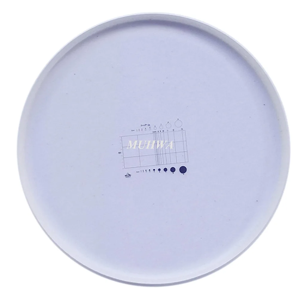
This article examines the optical design, calibration methodology, and measurement principles of net-reticle eyepiece micrometers, with specific reference to the C2 pattern — a 19 mm diameter glass disc inscribed with a rectangular grid of defined geometry, widely used in biological and materials science laboratories for morphometric quantification.
1. Optical Principle: The Intermediate Image Plane
In a compound microscope, the objective lens projects a real, magnified intermediate image at a fixed plane within the eyepiece tube. This plane — the field stop of the eyepiece — is precisely where the graticule is seated. Because both the specimen image and the graticule pattern are in sharp focus simultaneously, the scale appears superimposed on the specimen regardless of focus adjustments to the objective.
This principle has two practical implications. First, the graticule divisions represent arbitrary units (AU) that must be calibrated for each objective lens, because the magnification — and therefore the real-world distance corresponding to each division — differs at 10×, 40×, and 100×. Second, the graticule must be oriented such that its inscribed surface faces downward (toward the objective), placing it exactly at the focal plane. Installing the graticule upside down produces a blurred scale that cannot be brought into simultaneous focus with the specimen.
2. The C2 Net-Reticle Design

The C2 (√2ⁿ) Net-Shaped Eyepiece Micrometer is a 19 mm diameter optical glass disc engraved with a precision measurement grid. Unlike simple linear graticules, the C2 pattern integrates three measurement functions — particle counting, linear dimensioning, and particle sizing — into a single reticle. The engraved pattern is defined by the formula D = √2ⁿ × L/200, which governs the graduated scale of the right-half measurement zone and the diameter of the comparison circles, making the C2 an exceptionally versatile tool for quantitative microscopy in environmental monitoring, textile fiber inspection, public health, and industrial hygiene laboratories.
Its defining geometric characteristics are:
- 2:1 Rectangular Grid with 0-Line Partition: The central rectangle has length L and width L/2. A bold vertical 0-line bisects the rectangle into two equal squares. The left square is subdivided into a 3×2 grid (three vertical × two horizontal strips), forming six small rectangles for systematic particle counting within a defined area. The right square is divided into three horizontal strips, with unequally-spaced vertical graduations labeled 4, 6, 8, 9, 10, 11, 12, and 13. The distance from the 0-line to each graduation line follows the formula D = √2ⁿ × L/200, where n is the graduation index. This √2ⁿ geometric progression provides logarithmic measurement resolution, with finer spacing at small distances and coarser spacing at larger distances — well-suited for the wide size range of airborne particulates and textile fibers.
- Comparison Circles for Particle Sizing (n = 1 through 9): Above the rectangular grid, nine hollow circles of increasing diameter are arranged, each labeled with its index n (1–9). Below the grid, nine corresponding solid circles mirror the same diameters. The diameter D of circle n is given by D = √2ⁿ × L/200, which also equals the distance from the 0-line to the n-th graduation line in the right square. This dual calibration allows the operator to rapidly estimate particle diameters by visual comparison with the nearest hollow or solid circle, and to verify the measurement against the graduation scale in the right square. For example, circle 4 has a diameter equal to the distance from the 0-line to the graduation marked "4"; circle 5 equals the distance to the implicit graduation marked "5" (typically unlabeled); and so on through circle 9.
- 10-Aliquot Scale on the Left Edge: The extreme left margin of the grid is marked with 10 equally-spaced graduations, each representing L/20 of the total grid length. These aliquots provide a linear reference scale for quick one-dimensional length estimation without repositioning the specimen.
The C2 pattern is optimized for combined applications: the left 6-cell grid supports colony enumeration and particle density estimation (count per calibrated area); the right √2ⁿ-graduated scale supports precise linear measurement of fibers, dust particles, and biological structures; and the hollow/solid circle pairs enable rapid particle sizing by visual comparison — particularly valuable in occupational hygiene for asbestos fiber counting, in textile laboratories for fiber diameter measurement, and in environmental monitoring for particulate matter characterization.
3. Calibration Procedure: Graticule Units to Micrometers
The single most important practical skill in eyepiece micrometry is calibration against a certified stage micrometer. A stage micrometer is a microscope slide bearing a precision-etched scale of known length — typically 1 mm or 2 mm total, subdivided into 10 µm increments. The calibration protocol is:
- Insert the C2 graticule into the eyepiece, engraved surface downward. Rotate the eyepiece focusing ring until the grid lines are sharp.
- Place the stage micrometer on the microscope stage and focus using the 10× objective.
- Align the scales: Rotate the eyepiece so that the C2 grid lines are parallel to the stage micrometer scale. Translate the stage micrometer until the zero marks of both scales coincide.
- Determine the calibration factor: Count how many stage micrometer divisions (of known length) correspond to a known number of C2 graticule divisions. Calculate: Calibration factor (µm/division) = (number of stage µm) / (number of graticule divisions).
- Repeat for each objective: The calibration factor is objective-specific. Record the factor for 10×, 40×, and 100× objectives in a laboratory log. A typical calibration for a 10× objective yields approximately 10 µm per small graticule division; at 40×, approximately 2.5 µm; at 100× (oil immersion), approximately 1 µm.
Common calibration errors: Parallax — the apparent shift between the graticule and the stage micrometer scales when the observer's eye position changes — produces systematic measurement error. This is minimized by ensuring both scales are in the same focal plane and by viewing perpendicular to the eyepiece. A second common error is neglecting to recalibrate after changing objectives or after replacing the graticule in a different eyepiece position.
4. Measurement Applications
4.1 Linear Measurement
With the C2 grid calibrated, any structure aligned with the grid lines can be measured directly. For example, if the calibration factor at 40× is 2.5 µm per small division, a cell spanning 6 divisions measures 15 µm in diameter. The 10-aliquot subdivision provides sufficient resolution for distinguishing structures in the 5–500 µm range — covering typical eukaryotic cells, tissue features, fibers, and particulate contaminants.
4.2 Areal Measurement and Particle Counting
The rectangular net defines a known calibrated area. By counting objects (cells, colonies, particles, inclusions) that fall within the grid boundary and applying the appropriate counting rules (e.g., count objects touching the top and left borders; exclude those touching the bottom and right borders to avoid double-counting), the areal density is calculated as count / calibrated area. For particulate analysis or quality control applications where the grid area is calibrated in mm², this provides a direct count-per-unit-area metric without image capture or software processing.
4.3 Microscope Camera Calibration
When a microscope camera is attached, the C2 graticule serves as a calibration reference for digital image analysis. By capturing an image of the graticule at each objective, the pixel-to-micrometer conversion factor can be established for image analysis software (ImageJ, Fiji, NIS-Elements). This is particularly valuable in multi-user facilities where different cameras or software configurations may be in use — the graticule provides a hardware-based, software-independent calibration standard.
5. Material and Optical Quality Specifications
The metrological accuracy of an eyepiece micrometer depends on the optical quality of the glass substrate and the precision of the inscribed pattern. Key specifications include:
- Substrate material: Optical-grade borosilicate glass (refractive index ~1.52), selected for dimensional stability across laboratory temperature ranges (15–35°C). Thermal expansion of borosilicate glass (~3.3 × 10⁻⁶ /K) produces negligible scale error (~0.07% over a 20°C temperature swing).
- Inscription method: Chrome-deposited or acid-etched pattern with line width typically 2–5 µm. The lines must be sufficiently fine to provide a sharp reference edge without obscuring specimen detail, yet durable enough to withstand routine cleaning.
- Diameter: 19 mm — the standard eyepiece graticule diameter for most research-grade microscopes with 10×/20 mm or 10×/22 mm field number eyepieces. Compatibility should be verified against the specific eyepiece model; some manufacturers (Leica, Zeiss) use 21 mm or 26 mm formats.
6. Comparison with Alternative Measurement Methods
| Eyepiece Graticule (C2) | Stage Micrometer (Direct) | Digital Image Analysis | |
|---|---|---|---|
| Measurement type | Length, area, count (via overlay) | Length only (stage scale) | All parameters (post-capture) |
| Real-time? | Yes — measurement during observation | Yes | No — requires image capture and processing |
| Requires software? | No | No | Yes (ImageJ, Fiji, proprietary) |
| Accuracy | ±1–2% (after calibration) | ±0.1–0.5% (certified standard) | ±1–5% (dependent on calibration and segmentation) |
| Speed | Instant — measurement during live viewing | Moderate — repositioning required | Slow — capture, transfer, analyze workflow |
| Best application | Routine lab measurements, teaching, rapid QC screening | Calibration of graticules and digital systems | High-throughput batch analysis, publication-quality documentation |
The eyepiece graticule occupies a persistent and justified niche in modern microscopy: it is the only method that provides instant, real-time calibrated measurements during live observation, without interrupting the visual workflow for image capture. In teaching laboratories — where the pedagogical value of direct observation is irreplaceable — the graticule teaches students the relationship between the visual field and quantitative measurement in a way that software-based analysis cannot replicate.
7. Practical Recommendations
- Calibrate upon installation and verify periodically. Calibration should be repeated when objectives are changed, when the graticule is removed and reinserted, and at minimum every 6 months as part of routine microscope maintenance.
- Record calibration factors in a dedicated laboratory log or on a label affixed to the microscope body. Include the date, objective magnification, and calibration factor in µm/division.
- Clean with care. The graticule surface is precision-engraved. Use lens tissue moistened with 70% ethanol or isopropanol. Never apply dry tissue to a dry graticule — this abrades the inscribed pattern. Compressed air is preferred for routine dust removal.
- Verify against a certified standard annually. Stage micrometers with NIST-traceable or equivalent certification provide the reference standard against which graticule calibration is validated.
MUHWA C2 Net-Shaped Eyepiece Micrometer: 19 mm diameter optical glass graticule with 10-aliquot 2:1 rectangular grid pattern, compatible with standard 10×/20 mm eyepieces on major microscope brands (Olympus, Nikon, Zeiss, Leica, Motic). Available at muhwa-tech.com. Wholesale pricing available for educational and multi-user laboratory procurement.
This technical reference is provided for educational purposes. For applications requiring ISO/IEC 17025-accredited calibration, a certified stage micrometer with documented traceability to national metrology standards (NIST, PTB, NIM) is required. Eyepiece graticule calibration is only as accurate as the stage micrometer standard against which it is measured.
#microscope micrometer #eyepiece graticule #C2 reticle #microscope calibration #stage micrometer #cell measurement #morphometry #microscopy accessories #laboratory techniques #optical metrology
Comments
Post a Comment